Research at the Biomechanical Orthopedics Laboratory is divided in 4 areas: joint biomechanics, mechanobiology, tissue engineering, and drug delivery systems.
Joint Biomechanics
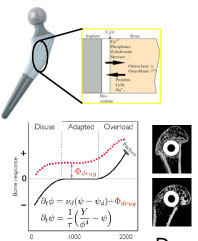
The research activities of the Joint Biomechanics Group consist in the development of theoretical, numerical, and experimental models to improve the clinical outcome of the treatement of the main humain joints: hip, knee, shoulder, ankle. One of our project consist in measuring the gap and micromotions at the bone-implant interface. The measurement is based on micro-CT imging coupled to a loading device.
Mechanobiology
Biomechanical results obtained in joints allow us to obtain boudary conditions which can be used as input for mechanobiology experiments. We are particularly interested to identify the different phenomena which lead to an implant loosening when its mechanical environment has been characterized. For exemple, we found that a short term duration mechanical stimulus applied on fresh human bone induced an increase RANKL/OPG ratio, leading then to an increase in bone resorption. This information was then used to design a drug delivery system by proposing to control the osteoclast in delivering bisphosphonate.
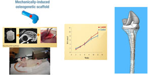
We are also interested to evaluate osteogenic mechanical stimulation in scaffold by evaluating the effect of fluid-induced shear stress on cells through scaffold deformation. This kind of information is then used in the design of bone scaffold.
![]()
Tissue engineering
The originality of our research in tissue engineering relies on two aspects. First we propose to use biomechanical stimulus to induce tissue formation in scaffold after its implantation and/or second, we use fetal cells seeded in the scaffold as a general cell source. We use this strategy specifically for bone and cartilage tissues.
Biomechanical stimulation
Based on biomechanical analysis, a composite scaffold (PLA and calcium-phosphate particles) has been developed in collaboration with the LTC (Dr. P. Bourban). This scaffold, with morphology and mechanical properties close to natural trabecular bone was implanted in rat femoral condyles. One leg of the rat was specifically loaded with a custom-made device while the other leg was kept as control. We observed a positive effect of the loading on the amount of bone formed in the loaded scaffold. The mechanical loading renders then the scaffold more osteoinductive and we propose to use this approach for specific loaded situation such as in revision of total knee arthroplasty.
Fetal cell therapy
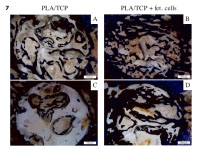
Fetal cells obtained from specific tissues present a high potential to generate cell bank. In particular for bone and cartilage, fetal cells have been characterized and several cell banks have been obtained in collaboration with the Unit Cell Therapy at the CHUV (Prof L. Applegate). The use of these cells directly seeded in the bone scaffold greatly increase the amount of bone formed.
Drug delivery systems
The presence of a fibrous tissue around orthopedic implants may lead to aseptic loosening, the main cause of implant failure. Tissue differentiation around implant (fibrous tissue instead of mineralized bone) is then a major parameter in the outcome success of an orthopedic implant. In this project, we propose to use the orthopedic implant as a drug delivery system in order to control the peri-implant tissue differentiation.
Specifically, by adding a bisphosphonate (Zoledronate) on an implant, we showed that not only a higher bone density was observed around the implant, but also its mechanical anchorage was greatly increased.
In parallel to the passvie delivery of drug, we also work on a smart delivery using mechanical stimulation as a release trigger.